
Scientists at Vilnius University (VU), together with partners in Hungary, are developing a new high-resolution optical measurement methodology based on the autofluorescence of algal cells and its correlation with their physiological state. This research could enable the development of a biosensor that uses algal health status to help detect pollution in bodies of water.
On 13–14 November, researchers from the Biological Research Centre of the Hungarian Research Network (HUN-REN) in Szeged visited the Faculty of Physics and the Life Sciences Centre at Vilnius University to discuss the results. According to VU researchers, the project is fostering and strengthening interdisciplinary collaboration in the development of innovative methods for monitoring environmental pollution.
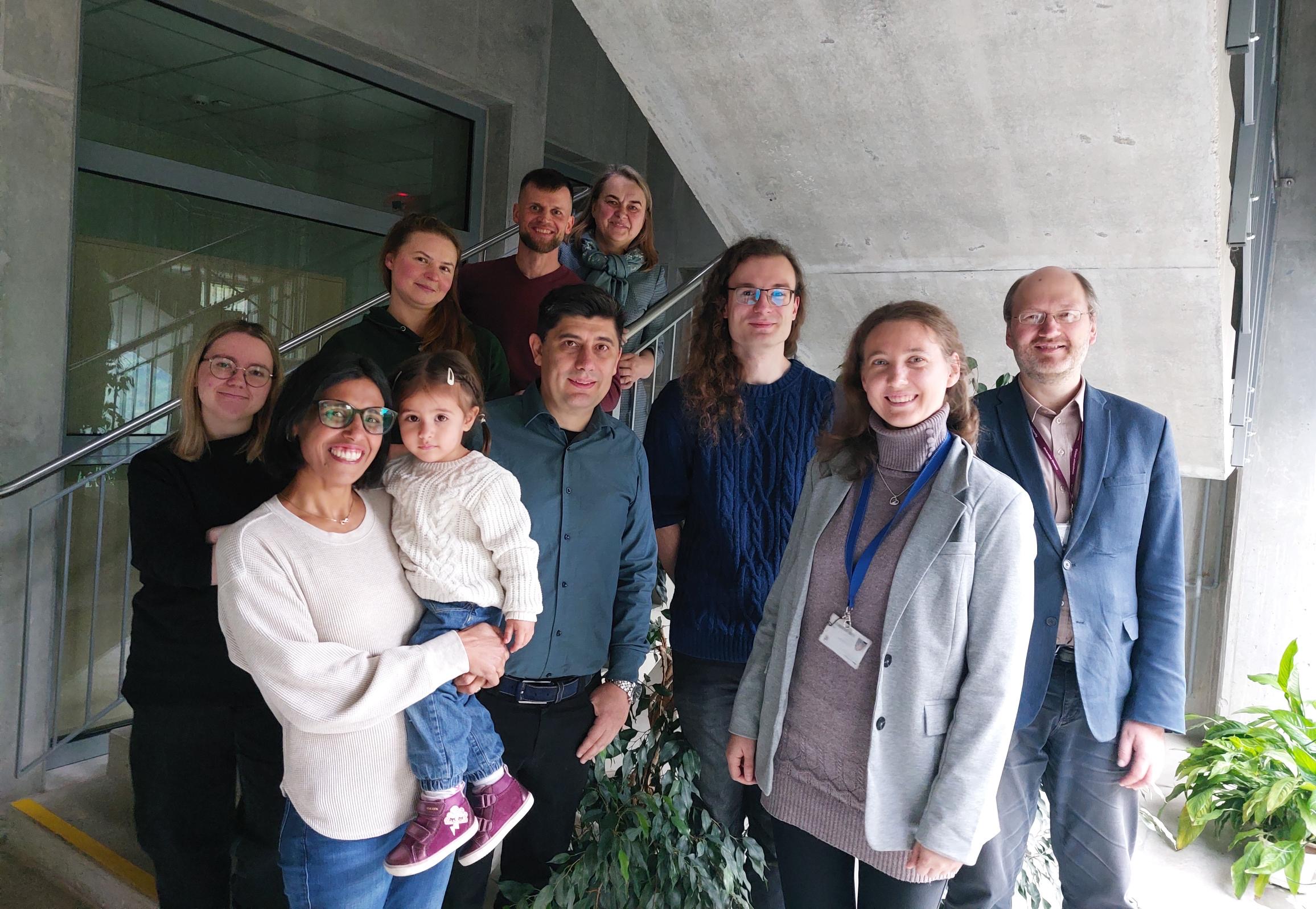
Group photo of guests and VU researchers. From left: Dr Parveen Akhtar next to Dr Petar Lambrev. Behind them: PhD student Aušrinė Navickaitė, Dr Indrė Lapeikaitė, PhD student Mykolas Mačiulis, and Prof. Vilma Kisnierienė. On the right: Prof. Saulius Bagdonas, Dr Agnė Kalnaitytė-Vengelienė, and Dr Vilmantas Pupkis.
A team of scientists from the Advanced Biomedical Photonics Group at the Laser Research Centre of the Faculty of Physics at Vilnius University and the Plant Cell Biophysics Research Group at the Department of Neurobiology and Biophysics at the Institute of Biosciences complement one another’s unique expertise. By combining optical and electrophysiological methods, the researchers examine freshwater algae subjected to photo-oxidative stress from multiple perspectives.
The methodology developed by the international research team links structural changes inside cells to photosynthetic signals that can be recorded remotely. “We study how photooxidative stress from different sources affects the vital functions and microstructure of algae under varying light conditions. By combining our expertise, we examine dynamic structural changes in living cells and their chloroplasts using optical spectroscopy, fluorescence microscopy and nonlinear polarimetric microscopy. This combination of methods, which is only beginning to be applied to algal cells, could form the basis for remote monitoring of signal changes, for example using drones,” says Prof. Saulius Bagdonas from the Faculty of Physics at Vilnius University, who leads the project’s research team in Lithuania. The research in Hungary is led by his colleague, Dr Petar Lambrev.
In addition to direct studies on algae, the project also includes fundamental research aimed at identifying potential ultra-sensitive biosensor components at the molecular level. During visits to Szeged, kinetic spectroscopy measurements are carried out using equipment at the HUN-REN Biological Research Centre and the Extreme Light Infrastructure Attosecond Light Pulse Source (ELI-ALPS).
“We analyse the behaviour of meso-tetra-(4-sulfonatophenyl)-porphyrin (TPPS4) molecules. In aqueous solution, these molecules form J-aggregates – large assemblies that efficiently transfer energy and are important for modelling energetic processes in photosynthesis. These aggregates are chiral, which makes them sensitive to light and magnetic fields. During our visits to Szeged, we looked for optimal conditions and sample-preparation protocols to form the largest possible TPPS4 aggregates with the highest degree of chirality. The greater the chirality (a symmetry property where an object does not coincide with its mirror image) and the larger the aggregate, the stronger and more sensitive their signal – an ideal feature when designing a highly sensitive biosensor,” explains Dr Agnė Kalnaitytė-Vengelienė.

Collaboration between Lithuanian and Hungarian researchers was initiated by Prof Virginijus Barzda of the University of Toronto, an alumnus of both Vilnius University and the University of Szeged. Together with early-career researchers, he carried out joint experiments during visits to Szeged as part of a project on nonlinear polarisation microscopy of dyes. This collaboration is continuing through new projects.