

Ya-Chi Chen and Loid Elizabeth Urtecho Navas, master's students in Chemical Engineering at the National Taiwan University of Science and Technology, spent a month at Vilnius University (VU) Faculty of Physics' Institute of Chemical Physics. Here, they investigated the potential of nanomaterials developed by their research group in Taiwan to improve Raman spectroscopy techniques.

From left: Loid Elizabeth Urtecho Navas and Ya-Chi Chen. Credit: Patricija Strumilaitė
Originally from Taiwan and Honduras, respectively, both students are members of Prof. Wei-Hung Chiang's research group. Their visit to Vilnius was part of a research collaboration between the two universities.
The collaboration began with a shared interest between the two research teams. While Prof. Chiang's group specialises in applying microplasma technology for nanomaterial synthesis, Prof. Valdas Šablinskas's team at VU offers expertise in Raman spectroscopy, diverse measurement capabilities, and experience in biomedical sensing research.
Exploring Nanomaterials Using Different Laser Wavelengths
The students' research focuses on gold- and silver- based nanomaterials synthesised using microplasma technology and their potential applications in surface-enhanced Raman spectroscopy (SERS).
SERS is a highly sensitive analytical method in which metal nanoparticles of a certain size significantly amplify the usually weak Raman spectroscopic signals. This makes it possible to detect extremely small quantities of substances under study. This method has been studied for decades and appears to hold promise for chemical analysis and biomedical research, as it produces strong and stable spectral signals. However, it remains unclear how to achieve reproducibility in experiments using this method. This would allow the method to be applied more widely in practice.
At VU, the students used Raman spectrometers with various lasers across a wide range of laser radiation wavelengths – from ultraviolet to infrared.
"In Taiwan, we design and synthesise the nanomaterials, while at Vilnius University, we carry out more in-depth SERS analysis," Urtecho explained.
Both students are happy to have the opportunity to exchange knowledge with VU researchers.
"Working closely with the researchers allowed us to better understand the spectral interpretation and the experimental design," she said.
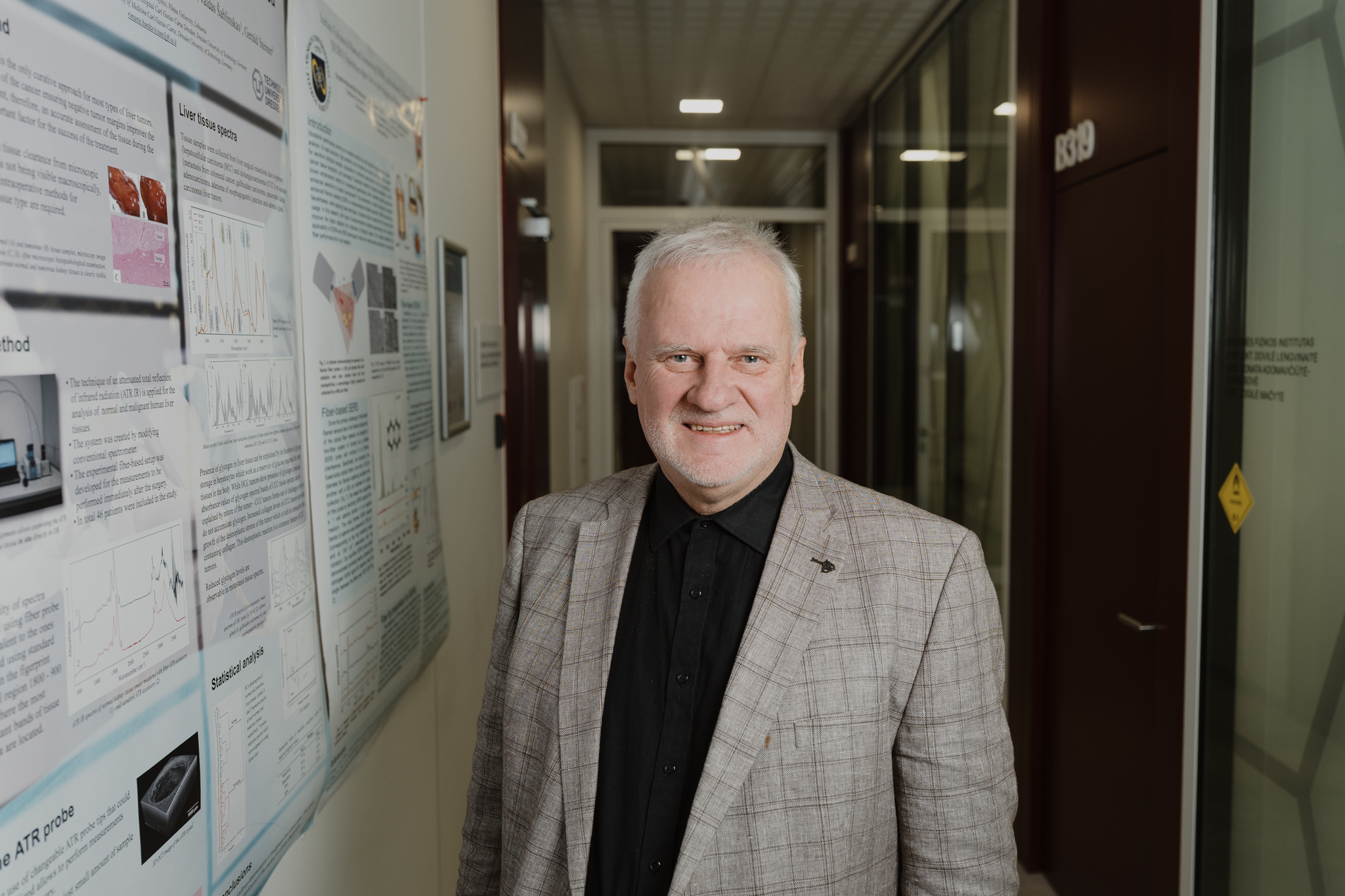
Prof. Valdas Šablinskas. Credit: Ugnius Bagdonavičius
Small Experimental Changes, New Perspectives
In Raman spectroscopy, the choice of substrate and careful sample handling can have a significant impact on the quality of the resulting spectra.
In Taiwan, their laboratory primarily uses silicon wafers as nanoparticle substrates. This approach allows researchers to optimise synthesis conditions and to characterise nanoparticles. At VU, conventional aluminium foil is used as a substrate, which allows researchers to reduce fluorescence background and to improve the signal-to-noise ratio in Raman and SERS measurements.
For Chen, another memorable aspect was the care taken by VU researchers to prevent contamination, including changing gloves when necessary and carefully cleaning the equipment and workspaces.
"Even a small amount of contamination can affect the experimental results," she observed.
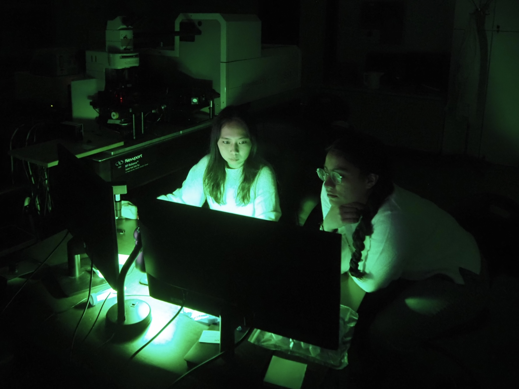 Chen and Urtecho examining measurement signals on a computer screen in the laboratory. Photo from personal archive
Research That Continues
Although the month-long exchange has come to an end, the research continues. The students’ plan is to analyse the measurements obtained in Vilnius along with their earlier results in Taiwan and use the findings to guide further material optimisation and the experimental design.
For now, Prof. Chiang's research group is testing the SERS signal-enhancing properties of its nanomaterials using commercial chemical compounds. This work forms part of the broader VU–Taiwan research collaboration aimed at developing biosensors for cancer diagnostics. Reaching the application stage, however, will require further testing and validation of the method using biomedical samples.
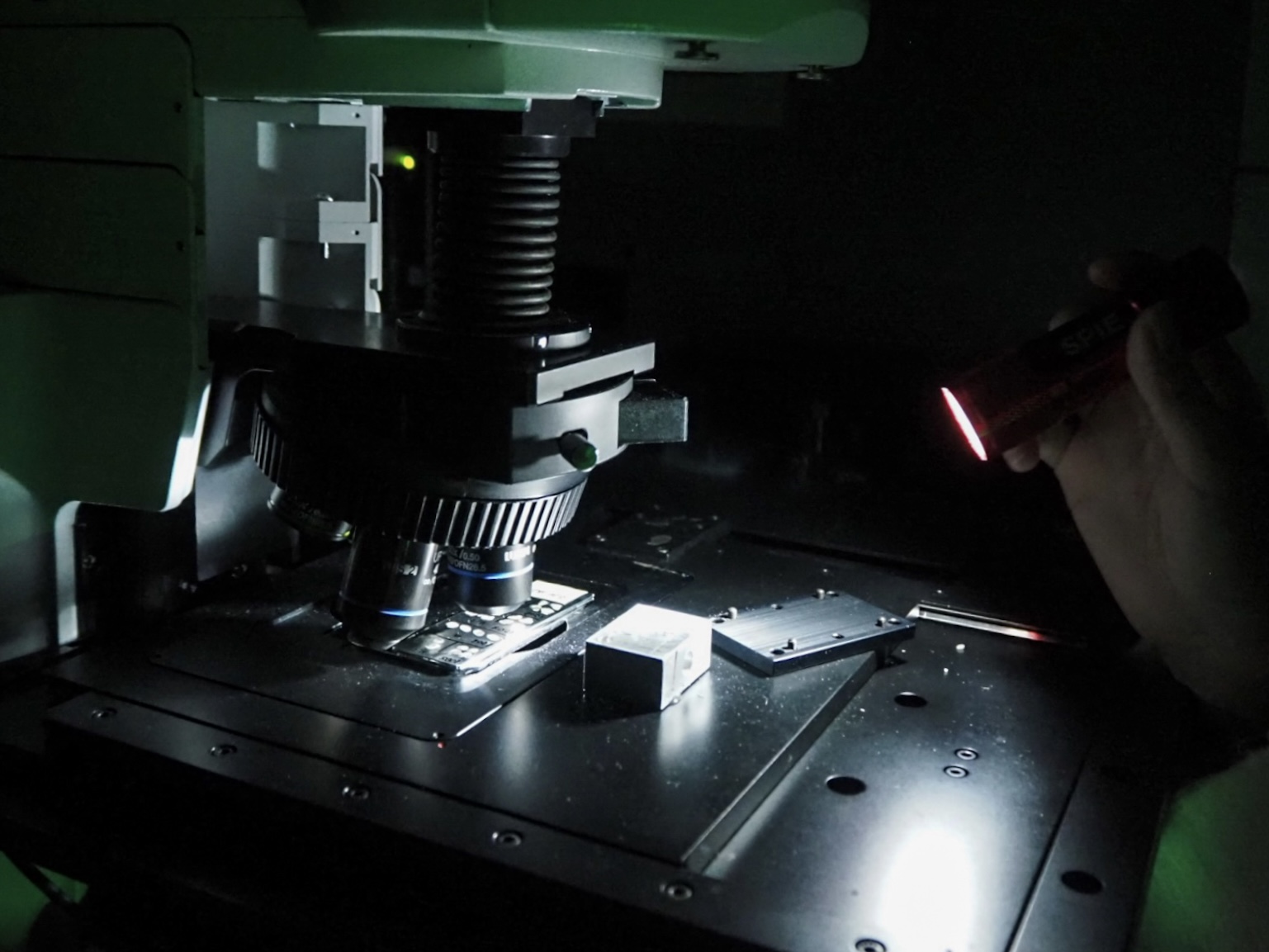
A sample positioned under the microscope for spectroscopic measurements. Photo from personal archive
For Chen and Urtecho, the exchange offered much more than additional experimental data. It was also an opportunity to learn from another research team and gain valuable experience that they can apply to their future research.